Biology: Respiratory System Diagram and Breathing

Breathing is something you do about twenty thousand times a day without thinking, yet the system behind it is a precise piece of engineering. Air must be cleaned, warmed, and delivered to tiny sacs where oxygen can enter the blood and carbon dioxide can leave it, and the whole process must run continuously for a lifetime. The respiratory system includes the airways, the lungs, and the muscles that move air in and out, and a diagram of it is a common exam question. This guide explains each part, how breathing works, and how gas exchange happens at the alveoli, so you can draw, label, and describe the system with confidence.
The Airways: From Nose to Lungs
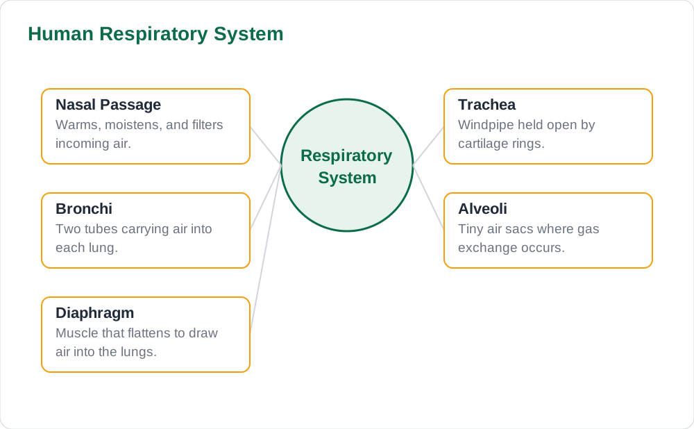
Air enters through the nose, where hairs and mucus trap dust and microbes, and where the air is warmed and moistened before it goes deeper. It then passes down the trachea, or windpipe, a wide tube held open by rings of cartilage so it never collapses, even when pressure changes during breathing. The trachea divides into two bronchi, one for each lung, and each bronchus divides again and again into smaller tubes called bronchioles, like branches of a tree. This branching structure carries air deep into the lungs while keeping the passages open and protected at every level.
The lining of the airways is important too, because it is covered with tiny hair-like cilia and a layer of mucus that traps dust and germs. The cilia beat in waves to sweep the mucus upwards towards the throat, where it is swallowed, in a cleaning system sometimes called the mucus escalator. Smoking damages these cilia, which is why smokers develop a persistent cough as mucus builds up. In a diagram, label the nose, trachea, bronchi, and bronchioles in order, and remember that cartilage rings belong to the trachea, not to the tiny bronchioles, which are too small to need them.
The Alveoli: Where Gas Exchange Happens
At the end of the bronchioles sit the alveoli, millions of tiny air sacs where gas exchange actually takes place. Each alveolus is surrounded by a dense network of blood capillaries, and both the alveolus wall and the capillary wall are only one cell thick, so gases diffuse across a very short distance. Oxygen diffuses from the air in the alveolus into the blood, where it binds to haemoglobin in red blood cells, and carbon dioxide diffuses the other way, from the blood into the alveolus, to be breathed out. This exchange happens by diffusion, moving from high concentration to low concentration, with no energy needed.
The alveoli are superbly adapted for their job, and examiners expect students to list these adaptations. They provide a huge total surface area, because there are millions of them, and their walls are thin and moist, which speeds diffusion. They also have a rich blood supply that constantly brings carbon dioxide and carries oxygen away, keeping the concentration gradients steep. A good exam answer names at least three adaptations and links each one to faster diffusion, rather than just listing them. Remember that gas exchange happens in the alveoli, not in the trachea or bronchi, which are only transport tubes.
Breathing In and Breathing Out
Breathing is driven by changes in the volume and pressure of the chest, and two sets of muscles control it: the diaphragm and the intercostal muscles between the ribs. When you breathe in, the diaphragm contracts and flattens downwards, and the intercostal muscles contract to pull the ribcage up and out. The chest volume increases, the air pressure inside the lungs falls below the outside air pressure, and air rushes in. When you breathe out, these muscles relax, the diaphragm domes back up, the ribcage falls, the chest volume decreases, and air is pushed out. This is why breathing in is an active process and normal breathing out is mostly passive.
Students often confuse breathing with respiration, so keep the distinction clear. Breathing is the physical movement of air in and out of the lungs, while respiration is the chemical process inside cells that releases energy from glucose. Breathing supplies the oxygen that respiration needs and removes the carbon dioxide it produces, but the two are not the same thing. During exercise, breathing rate and depth both increase, because active muscles need more oxygen and produce more carbon dioxide, and the brain detects the rising carbon dioxide level and signals faster breathing to restore balance.
How Gas Exchange Stays Efficient
Gas exchange stays efficient because the body constantly maintains steep concentration gradients at the alveoli. Fresh air arriving with each breath keeps oxygen high in the alveoli, while blood arriving from the body is low in oxygen, so oxygen diffuses rapidly into the blood. At the same time, blood arriving is high in carbon dioxide, while alveolar air is low in carbon dioxide, so carbon dioxide diffuses out just as quickly. The heart keeps blood flowing through the lung capillaries, so the gradients never flatten, and this continuous flow is as important as the thin walls and large surface area.
The same principles explain gas exchange in other organisms and in other organs. In the tissues, the gradients reverse: oxygen diffuses from the blood into the cells, and carbon dioxide diffuses from the cells into the blood, because respiring cells use oxygen and produce carbon dioxide. A student who understands gradients can answer questions about lungs, tissues, and even gills in fish, because the same rule, diffusion down a concentration gradient across a thin, moist surface, applies everywhere. Always mention the gradient in a gas exchange answer, because it is the reason diffusion happens at all.
Drawing the Diagram and Common Mistakes
Draw the respiratory system with the trachea in the centre, the two bronchi branching into the lungs, and the diaphragm as a dome shape below them. Show the lungs as two large organs on either side, and label the alveoli as tiny sacs at the ends of the bronchioles, often drawn as a small cluster in a magnified inset. Label the ribs and intercostal muscles only if the question asks about the breathing mechanism, but always label the diaphragm, because it is the main breathing muscle. Keep leader lines straight and outside the diagram, and add arrows showing air moving in and out to make the direction clear.
Common mistakes include labelling the trachea as the oesophagus, drawing the diaphragm above the lungs, and saying that oxygen is breathed in and carbon dioxide is breathed out as if inhaled air were pure oxygen. Inhaled air is mostly nitrogen, with about one fifth oxygen, and exhaled air still contains plenty of nitrogen and some oxygen, just with more carbon dioxide and water vapour. For more biology revision guides and solved notes, explore Solved Notes, browse the full collection on Books, and read other study articles on the Blog to finish your revision of breathing and respiration.
AI Help: Muse AI
Muse AI is Meta's personal AI assistant, and it can help you learn breathing by explaining the in-and-out mechanism step by step, or by quizzing you on alveolar adaptations. You can ask it to describe what the diaphragm does during inhalation, or to test you on the difference between breathing and respiration. Muse AI does not give official STBB answers — always verify against your STBB book and your teacher. Always verify against your Sindh Textbook Board book and your teacher before using anything in classwork.
Join Muse with invite code LTGSWD — in the Muse app, go to Settings > Redeem token and enter the code. Disclosure: LTGSWD is the author's Muse invite code.
5 key points you must know
- Air travels nose, trachea, bronchi, bronchioles, then alveoli, in that order.
- Alveoli have thin moist walls, huge surface area, and a rich blood supply for fast diffusion.
- Breathing in: diaphragm flattens, ribs move up and out, chest volume rises, air rushes in.
- Breathing is air movement; respiration is energy release from glucose inside cells.
- Inhaled air is mostly nitrogen, not pure oxygen; exhaled air still contains some oxygen.
Frequently asked questions
What happens to the diaphragm when you breathe in?
The diaphragm contracts and flattens downwards, increasing the volume of the chest. This lowers the air pressure inside the lungs below the outside pressure, so air rushes in. At the same time, the intercostal muscles contract to lift the ribcage up and out, adding further volume for a deep breath.
Why are alveoli good for gas exchange?
Alveoli have a huge total surface area because there are millions of them, walls only one cell thick, and a dense capillary network. Their walls are moist, which helps gases dissolve and diffuse quickly. The constant blood flow maintains steep concentration gradients, so oxygen keeps diffusing in and carbon dioxide keeps diffusing out.
What is the difference between breathing and respiration?
Breathing is the physical movement of air into and out of the lungs, driven by the diaphragm and rib muscles. Respiration is the chemical process in cells that releases energy from glucose, using oxygen and producing carbon dioxide. Breathing supplies oxygen for respiration and removes its carbon dioxide waste, but they are different processes.
Why does breathing rate increase during exercise?
Exercising muscles respire faster, using more oxygen and producing more carbon dioxide. The brain detects the rising carbon dioxide level in the blood and signals faster, deeper breathing. This brings in more oxygen and removes carbon dioxide more quickly, keeping the blood chemistry balanced during activity.
What is the job of cilia and mucus in the airways?
Mucus traps dust, pollen, and microbes from inhaled air, and cilia, tiny hairs lining the airways, beat in waves to sweep the mucus upwards to the throat, where it is swallowed. This cleaning system protects the delicate alveoli from particles and infection. Smoking damages cilia, so mucus builds up and causes a persistent cough.
Solved notes — preview free
These guides work best with the solved exercises. Preview the solved notes free on this website — 119 solved notes for Classes 6–10 in Sindhi, Urdu, English and Arabic medium. Or browse the full solved notes catalogue for your class and medium. New solved notes are added regularly — check the catalogue for coming-soon books.