Biology: Leaf Structure Diagram and Functions

A leaf looks like a simple flat green blade, but a cross-section under the microscope reveals a carefully organised structure in which every layer has a job. The leaf must capture light, exchange gases, move water in and food out, and avoid losing too much water, all at the same time. Each layer of the leaf is adapted for one of these tasks, and understanding the layers is the key to understanding photosynthesis itself. This guide walks through the leaf from top to bottom, explains what each layer does, and shows how to draw and label a leaf cross-section diagram that examiners will reward.
The Upper Layers: Cuticle and Epidermis
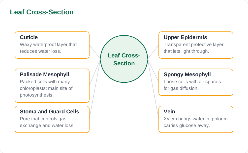
The top of the leaf is covered by the cuticle, a waxy, waterproof layer that reduces water loss by evaporation. Below it lies the upper epidermis, a single layer of thin, transparent cells with no chloroplasts, which protects the leaf while letting light pass through to the layers below. These two layers work as a team: the cuticle keeps water in, and the transparent epidermis lets light in, and neither layer blocks the other. A leaf in a hot, dry habitat usually has a thicker cuticle, which is a clear example of adaptation to the environment, and a good detail to mention in an exam answer about water conservation.
The epidermis also has a protective role against microbes and physical damage, because it forms a continuous skin over the leaf surface. Its cells fit tightly together with no gaps, so water cannot escape easily and pathogens cannot enter easily. When you draw a leaf cross-section, show the cuticle as a thin line on top and the upper epidermis as a single row of cells beneath it, and label both separately. Students often merge them into one layer, but examiners award separate marks for each, so drawing them as two distinct lines is worth the small extra effort.
The Palisade Layer: The Photosynthesis Workshop
Directly below the upper epidermis sits the palisade mesophyll, a layer of tall, tightly packed cells standing upright like a fence, and this is where most photosynthesis happens. Palisade cells contain large numbers of chloroplasts, and their upright shape lets light pass down between them while their close packing fits many chloroplasts near the top of the leaf, where light is strongest. This layer is the reason leaves are green on top and why the upper surface faces the sun: the plant places its light-capturing machinery exactly where the light arrives, which is efficient design by any standard.
The palisade layer is the most important layer to label correctly, because exam questions about where photosynthesis happens almost always expect this answer. Remember that photosynthesis also happens in the spongy layer below, but at a lower rate, because it has fewer chloroplasts and receives less light. When labelling, draw palisade cells as tall rectangles packed together, clearly different from the rounded, loosely arranged cells below them. The visual contrast between the two mesophyll layers is what makes a leaf diagram convincing, so do not draw both layers as identical rows of squares.
The Spongy Layer and Air Spaces
Below the palisade layer lies the spongy mesophyll, a layer of rounded cells arranged loosely with large air spaces between them. These air spaces are essential, because they let carbon dioxide diffuse easily to the palisade cells and let oxygen diffuse out, connecting the inside of the leaf to the outside air through the stomata. Spongy cells contain some chloroplasts, so they also photosynthesise, but their main contribution is gas movement rather than light capture. The loose arrangement also makes the leaf light and flexible, and it increases the internal surface area over which gases can diffuse.
The air spaces explain why a leaf floats and why it feels soft and spongy when pressed, and they are a favourite exam detail. A common mistake is to draw the spongy layer as tightly packed as the palisade layer, which removes the air spaces and the reason for the layer's name. Draw rounded cells with clear gaps between them, and label one gap as an air space if you have room. Linking structure to function here, air spaces for gas diffusion, shows the kind of understanding that moves an answer from average to excellent, because it proves you know why the layer looks the way it does.
Veins, Stomata, and Guard Cells
Running through the mesophyll are the veins, the leaf's transport system, each containing xylem and phloem. Xylem vessels bring water and dissolved minerals up from the roots to the leaf, supplying the water photosynthesis needs, while phloem tubes carry the glucose made in the leaf to other parts of the plant for growth or storage. Veins also support the leaf mechanically, acting like the ribs of an umbrella to hold the blade flat and spread out for maximum light capture. In a cross-section, draw a vein as a bundle in the middle of the mesophyll, and label xylem and phloem separately if the question asks for detail.
On the lower surface, the lower epidermis contains tiny pores called stomata, each surrounded by two guard cells. Guard cells control the opening and closing of the stoma: when they take in water and become turgid, the pore opens and gases exchange freely, and when they lose water, the pore closes to conserve water. Most stomata are on the lower surface, which is cooler and less exposed to direct sun, reducing water loss. This control is a balancing act between taking in carbon dioxide for photosynthesis and keeping water inside the leaf, and it is one of the most elegant adaptations in plant biology.
Drawing the Leaf Diagram and Revision Tips
Draw the leaf cross-section as a wide, flat rectangle with the layers stacked in order: cuticle, upper epidermis, palisade layer, spongy layer, lower epidermis, and stomata on the lower surface, with a vein in the middle. Make the palisade cells tall and packed and the spongy cells round and spaced, so the two layers are instantly different. Label each layer with a straight leader line, and put labels on both sides to avoid crowding. A neat, correctly ordered cross-section with six or more labels is one of the highest-scoring diagrams in plant biology, because it tests so many facts in one picture.
Revise by covering the labels and naming each layer from top to bottom, then stating one function for each. Ask yourself why the palisade layer is at the top, why stomata are mostly below, and why the cuticle is waxy, and answer each question with a function, not just a name. For more biology revision guides and solved notes, explore Solved Notes, browse the full collection on Books, and read other study articles on the Blog to complete your revision of plant structure and photosynthesis.
AI Help: Muse AI
Muse AI is Meta's personal AI assistant, and it can help you learn leaf structure by naming a layer and asking you to recall its function, like a flashcard quiz. You can ask it to walk through a cross-section from top to bottom, or to explain why guard cells open and close the stomata. Muse AI does not give official STBB answers — always verify against your STBB book and your teacher. Always verify against your Sindh Textbook Board book and your teacher before using anything in classwork.
Join Muse with invite code LTGSWD — in the Muse app, go to Settings > Redeem token and enter the code. Disclosure: LTGSWD is the author's Muse invite code.
5 key points you must know
- The cuticle is waxy and waterproof, reducing water loss from the leaf surface.
- The upper epidermis is transparent, letting light reach the palisade layer below.
- The palisade mesophyll has many chloroplasts and is the main site of photosynthesis.
- The spongy mesophyll has air spaces that allow gases to diffuse through the leaf.
- Stomata, controlled by guard cells, balance carbon dioxide intake against water loss.
Frequently asked questions
Why is the palisade layer the main site of photosynthesis?
Palisade cells are packed with chloroplasts and sit just below the transparent upper epidermis, where light is strongest. Their tall, upright shape lets light penetrate deeply while fitting many chloroplasts into the top of the leaf. The spongy layer below also photosynthesises, but it has fewer chloroplasts and receives less light, so its rate is lower.
How do guard cells open and close a stoma?
Guard cells surround each stoma, and when they absorb water they become turgid and curve apart, opening the pore so gases can exchange. When they lose water they become flaccid and close together, shutting the pore to conserve water. This mechanism lets the leaf balance carbon dioxide intake for photosynthesis against water loss by evaporation.
Why are most stomata on the lower surface of the leaf?
The lower surface is cooler and less exposed to direct sunlight than the upper surface, so less water evaporates through stomata there. Placing stomata below therefore reduces water loss while still allowing gas exchange. The upper surface, which faces the sun, is protected instead by the waxy cuticle and the epidermis.
What do xylem and phloem do in a leaf vein?
Xylem vessels bring water and dissolved minerals from the roots up to the leaf, supplying the water needed for photosynthesis. Phloem tubes carry glucose made in the leaf to other parts of the plant, such as growing shoots, roots, and storage organs. Veins also support the leaf blade, holding it flat and spread out to capture maximum light.
What is the function of the air spaces in the spongy mesophyll?
Air spaces let carbon dioxide diffuse easily from the stomata to the photosynthesising cells, and let oxygen diffuse out again. They increase the internal surface area for gas exchange and connect the inside of the leaf to the outside air. Without these spaces, gases would move slowly through solid tissue and photosynthesis would be much less efficient.
Solved notes — preview free
These guides work best with the solved exercises. Preview the solved notes free on this website — 119 solved notes for Classes 6–10 in Sindhi, Urdu, English and Arabic medium. Or browse the full solved notes catalogue for your class and medium. New solved notes are added regularly — check the catalogue for coming-soon books.