Biology: Human Heart Diagram and Blood Flow

The human heart is a muscular pump about the size of your fist, and its job is to push blood around the body day and night without ever stopping. Blood carries oxygen and food to every cell and collects waste products like carbon dioxide, so the heart's steady pumping is what keeps every organ alive. A heart diagram is a favourite exam question because it tests chambers, vessels, valves, and the direction of blood flow all at once. This guide explains the four chambers, the main blood vessels, the valves, and the double circulation, and shows how to draw and label a heart diagram that earns full marks.
The Four Chambers of the Heart
The heart is divided into four chambers: two atria at the top and two ventricles at the bottom, with a left side and a right side that never mix. The right atrium receives deoxygenated blood from the body, and the left atrium receives oxygenated blood from the lungs. Below them, the right ventricle pumps blood to the lungs, while the left ventricle pumps blood to the whole body. A wall called the septum separates the left and right sides, so oxygenated and deoxygenated blood stay apart, which is essential for efficient delivery of oxygen to the tissues.
The walls of the chambers are not all the same thickness, and this is a detail examiners love. The left ventricle has the thickest, most muscular wall, because it must pump blood all the way around the body, against resistance, with every beat. The right ventricle has a thinner wall, because it only pumps blood a short distance to the nearby lungs. The atria have the thinnest walls of all, because they only push blood a short way down into the ventricles. When you label a heart diagram, pointing out the thick left ventricle wall shows that you understand why the heart is shaped the way it is, not just what it is called.
The Main Blood Vessels Attached to the Heart
Five main vessels connect to the heart, and each one has a clear job. The vena cava brings deoxygenated blood from the body into the right atrium. The pulmonary artery carries deoxygenated blood from the right ventricle to the lungs, and it is the exception to the usual rule, because it is an artery carrying deoxygenated blood. The pulmonary vein brings oxygenated blood from the lungs into the left atrium, and it is the opposite exception, a vein carrying oxygenated blood. Finally, the aorta, the largest artery in the body, carries oxygenated blood from the left ventricle to the rest of the body.
A simple rule helps with most vessels: arteries carry blood away from the heart, and veins carry blood towards the heart. In most of the body, arteries carry oxygenated blood and veins carry deoxygenated blood, but the pulmonary vessels break this pattern because they serve the lungs, where blood is re-oxygenated. Remembering the two exceptions, pulmonary artery with deoxygenated blood and pulmonary vein with oxygenated blood, prevents the most common vessel mistake in exams. Label each vessel on your diagram with an arrow showing the direction of flow, because direction is often worth marks on its own.
Valves: The One-Way Doors
Valves are one-way doors inside the heart that stop blood flowing backwards, and they are essential because blood must move in a single direction through the chambers. Valves sit between each atrium and ventricle, and also at the exits of the ventricles into the pulmonary artery and the aorta. When a chamber contracts, its valve opens to let blood through, and when the chamber relaxes, the valve snaps shut to prevent backflow. The familiar lub-dub sound of the heartbeat is made by these valves closing, first the atrioventricular valves and then the semi-lunar valves at the artery exits.
Understanding valves helps explain why the heart is such an efficient pump. Without valves, blood would slosh backwards with every beat and never build up enough pressure to reach distant organs. In a diagram, valves are usually drawn as small flaps or lines at the chamber exits, and labelling them correctly shows extra understanding. Students sometimes forget valves entirely, so make them a fixed part of your drawing checklist: chambers, vessels, valves, arrows, and then labels. A diagram with valves labelled looks complete, while one without them looks unfinished to an examiner.
Blood Flow Step by Step: Double Circulation
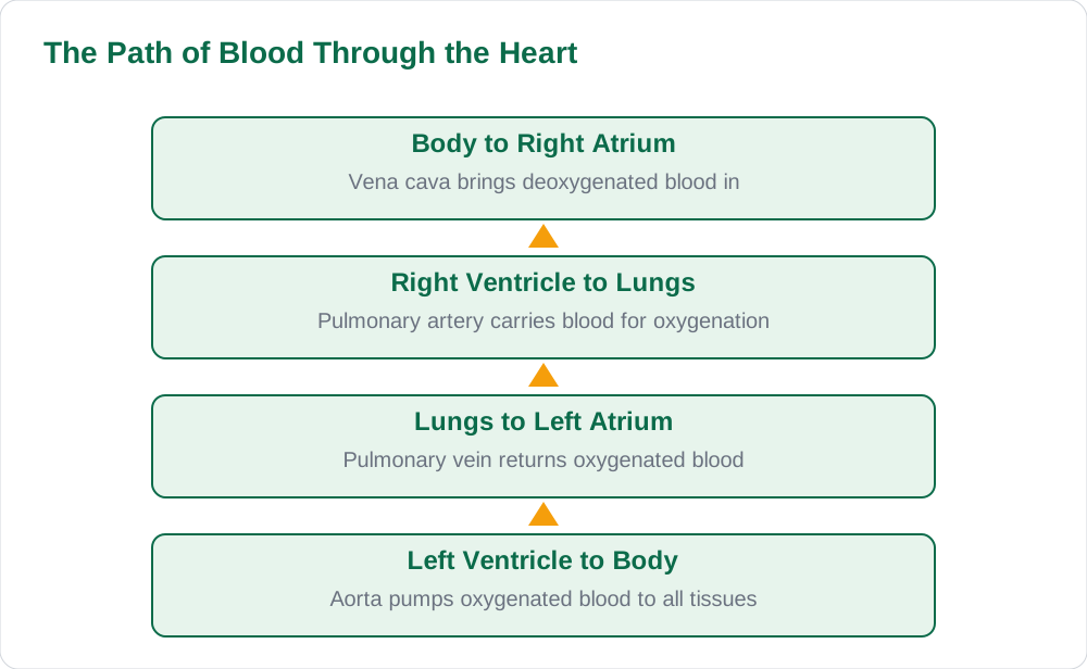
Blood flows through the heart in a fixed sequence that you should be able to recite in order. Deoxygenated blood from the body enters the right atrium through the vena cava, passes into the right ventricle, and is pumped through the pulmonary artery to the lungs. In the lungs, carbon dioxide is removed and oxygen is picked up, and the now oxygenated blood returns through the pulmonary vein into the left atrium. It then passes into the left ventricle, which pumps it through the aorta to the whole body, where cells take oxygen and release carbon dioxide, and the cycle begins again.
This is called double circulation because blood passes through the heart twice for each complete circuit of the body: once on its way to the lungs, and once on its way to the body. The pulmonary circuit carries blood between the heart and the lungs, while the systemic circuit carries blood between the heart and the rest of the body. Double circulation is efficient because blood is re-pressurised and re-oxygenated before it is sent around the body, so oxygen reaches the tissues quickly and at a good pressure. Mammals, including humans, need this efficient system because they are active, warm-blooded animals with high energy demands.
Drawing the Heart Diagram and Avoiding Mistakes
Draw the heart as a roughly cone-shaped organ with the pointed end, the apex, facing slightly to your left and downwards, which is how it sits in the chest. Mark the four chambers first, then add the vessels at the top, keeping the aorta as the thickest arch on the left side of the diagram. Add arrows to every vessel to show flow direction, and label valves where they sit between chambers and at artery exits. Keep labels outside the diagram with straight leader lines, and do not cross the lines. A heart diagram takes practice, so redraw it from memory three or four times before the exam until the chamber positions feel automatic.
The most common mistakes are swapping the left and right sides, forgetting the pulmonary exceptions, and leaving out arrows. Remember that the left side of the heart is drawn on the right side of the page, because you are looking at someone else's heart facing you, and examiners check this carefully. Also, never draw blood flowing backwards through a valve, and never label the aorta as a vein. For more biology revision guides and solved notes, explore Solved Notes, browse the full collection on Books, and read other study articles on the Blog to prepare thoroughly for your exams.
AI Help: Muse AI
Muse AI is Meta's personal AI assistant, and it can help you learn the heart by walking you through blood flow chamber by chamber, like a guided tour of the diagram. You can ask it to quiz you on which vessel carries which type of blood, or to explain double circulation in simple words. Muse AI does not give official STBB answers — always verify against your STBB book and your teacher. Always verify against your Sindh Textbook Board book and your teacher before using anything in classwork.
Join Muse with invite code LTGSWD — in the Muse app, go to Settings > Redeem token and enter the code. Disclosure: LTGSWD is the author's Muse invite code.
5 key points you must know
- The heart has four chambers: right and left atria above, right and left ventricles below.
- The septum keeps oxygenated and deoxygenated blood separate.
- The left ventricle wall is thickest because it pumps blood around the whole body.
- Pulmonary artery carries deoxygenated blood; pulmonary vein carries oxygenated blood.
- Double circulation means blood passes through the heart twice per full body circuit.
Frequently asked questions
Why is the left ventricle wall the thickest?
The left ventricle pumps oxygenated blood through the aorta to the entire body, which needs high pressure to reach distant tissues. Its thick muscular wall generates that pressure with every beat. The right ventricle only pumps blood a short distance to the lungs, so its wall is thinner, and the atria are thinnest because they only push blood down into the ventricles.
What is double circulation?
Double circulation means blood passes through the heart twice during one complete circuit of the body. The pulmonary circuit sends blood to the lungs for oxygenation, and the systemic circuit sends oxygenated blood to the rest of the body. This re-pressurises and re-oxygenates the blood, making oxygen delivery faster and more efficient than a single circulation.
Which vessels are the exceptions to the artery and vein rule?
The pulmonary artery is an artery that carries deoxygenated blood from the right ventricle to the lungs, and the pulmonary vein is a vein that carries oxygenated blood from the lungs to the left atrium. Everywhere else, arteries carry oxygenated blood away from the heart and veins carry deoxygenated blood towards it. Remembering these two exceptions avoids the most common vessel mistake.
What do heart valves do?
Valves are one-way doors that prevent blood from flowing backwards through the heart. They sit between the atria and ventricles and at the exits into the pulmonary artery and aorta. When a chamber contracts, the valve opens to let blood through, and when it relaxes, the valve closes, and this closing makes the lub-dub heartbeat sound.
Why is the left side of the heart drawn on the right of the page?
Because a heart diagram shows the heart as it appears in another person's chest facing you, so left and right are reversed from your own point of view. The left ventricle, which is the heart's left side, therefore appears on the right side of the page. Examiners check this carefully, so always label sides from the heart owner's perspective, not your own.
Solved notes — preview free
These guides work best with the solved exercises. Preview the solved notes free on this website — 119 solved notes for Classes 6–10 in Sindhi, Urdu, English and Arabic medium. Or browse the full solved notes catalogue for your class and medium. New solved notes are added regularly — check the catalogue for coming-soon books.