Biology: Digestive System Diagram From Mouth to Intestine
Digestion is the process that breaks large, insoluble food molecules into small, soluble ones that the blood can absorb and carry to every cell. Without it, the food we eat could never reach our tissues, no matter how much we ate. The human digestive system is a long tube, with helper organs attached, running from the mouth to the anus, and each section has a specialised job. This guide follows food through every stage, explains the enzymes and organs involved, and shows how to draw and label a digestive system diagram that covers the whole journey in the correct order.
The Mouth: Where Digestion Begins
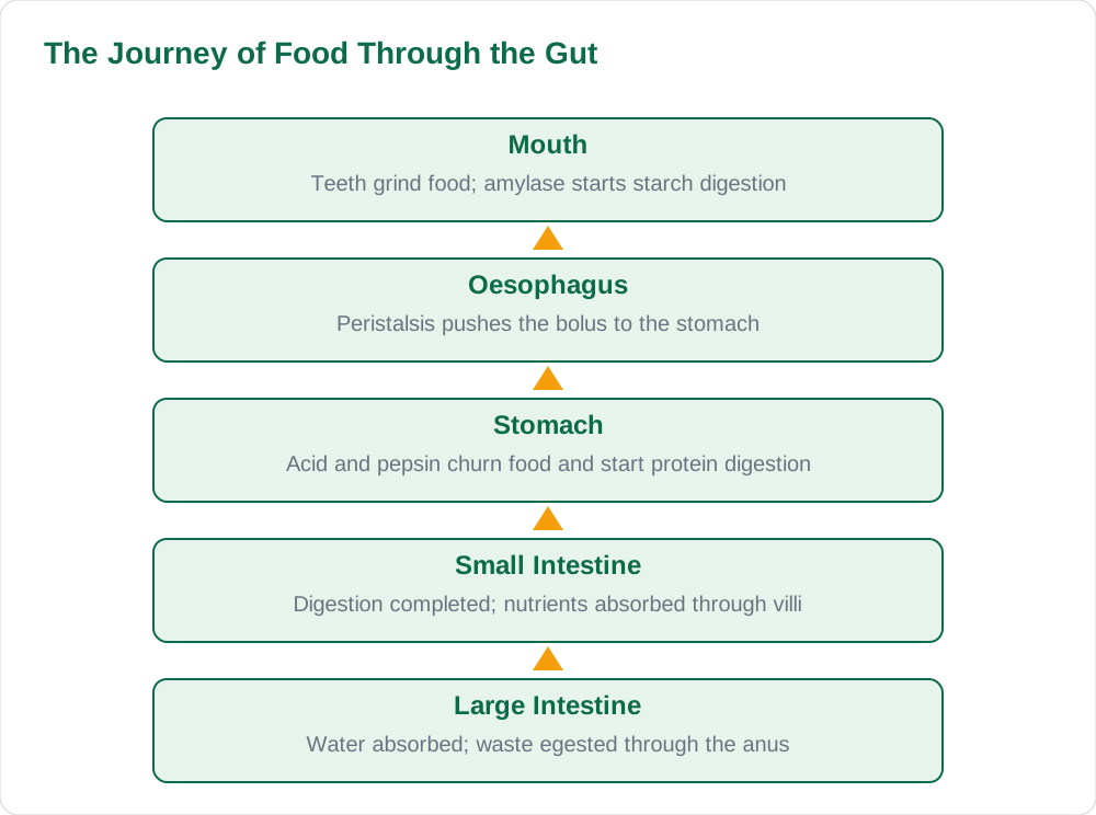
Digestion begins in the mouth, where two kinds of breakdown happen at once. Teeth carry out mechanical digestion, cutting, tearing, and grinding food into smaller pieces, which increases the surface area for enzymes to work on. At the same time, salivary glands release saliva, which contains the enzyme amylase, and amylase begins chemical digestion by breaking starch into smaller sugars. Saliva also moistens food and rolls it into a soft ball called a bolus, which is easy to swallow. Chewing well therefore matters for digestion, not just for comfort, because smaller pieces and early starch digestion give the rest of the system a head start.
The four types of teeth each have a role: incisors cut, canines tear, premolars crush, and molars grind. Humans have two sets of teeth in a lifetime, milk teeth in childhood and permanent teeth later, and keeping them clean protects the whole digestive process. After swallowing, the bolus travels down the oesophagus, a muscular tube that pushes food towards the stomach by wave-like contractions called peristalsis. Peristalsis continues through the whole gut, so food keeps moving even when you are upside down, and no digestion of note happens in the oesophagus itself, which is purely a transport tube.
The Stomach: Acid and Churning
In the stomach, food is churned by muscular walls and mixed with gastric juice, which contains hydrochloric acid and the enzyme pepsin. The acid kills many harmful microbes swallowed with food and provides the acidic conditions pepsin needs to work, while pepsin begins digesting proteins into smaller pieces. The stomach lining is protected from its own acid by a layer of mucus, which is why the stomach does not digest itself under normal conditions. Food stays in the stomach for a few hours, being churned into a thick, soupy mixture called chyme, which is then released in small amounts into the small intestine.
A ring of muscle at the stomach exit, the pyloric sphincter, controls this release so the small intestine is not overwhelmed. This is an important exam point: the stomach does not absorb much food itself, and its main jobs are storage, churning, acid protection, and the start of protein digestion. Students often write that digestion is completed in the stomach, which is wrong; the stomach only begins the process. The real completion of digestion and nearly all absorption happen further along, in the small intestine, which is the next and longest section of the journey.
The Small Intestine: Digestion Completed and Absorbed
The small intestine is where digestion is completed and where almost all absorption takes place, and it is long and folded to give a huge surface area. Three helper secretions arrive here: bile from the liver, stored in the gall bladder, which neutralises stomach acid and emulsifies fats into tiny droplets, and pancreatic juice from the pancreas, which contains enzymes for carbohydrates, proteins, and fats. Enzymes in the intestinal wall then finish the job, breaking the remaining pieces into glucose, amino acids, fatty acids, and glycerol, which are small and soluble enough to be absorbed into the blood.
Absorption happens through the villi, millions of tiny finger-like projections lining the small intestine wall. Each villus has a thin wall, a rich blood supply, and a large surface area, all adaptations that speed absorption. Glucose and amino acids pass into the blood capillaries in the villus and travel to the liver, while fatty acids and glycerol pass into the lacteal, a small lymph vessel. This is why the villi are the most labelled structure in digestive diagrams: their structure matches their function perfectly, and examiners expect students to explain at least two adaptations, such as thin walls and a good blood supply, for full marks.
The Large Intestine, Liver, and Pancreas
Whatever is left after the small intestine, mainly water, fibre, and undigested material, passes into the large intestine. Here, water is absorbed back into the body, which is why the remaining material becomes more solid as it travels along. Bacteria living in the large intestine also produce some vitamins, and the solid waste, faeces, is stored in the rectum before being removed through the anus, a process called egestion. Egestion is different from excretion, because faeces were never inside the body's cells; they are simply undigested material leaving the gut tube, and examiners often test this distinction.
The liver and pancreas deserve their own labels on any digestive diagram because they are essential helper organs. The liver makes bile, which emulsifies fats, and it also processes absorbed nutrients, stores glucose as glycogen, and breaks down harmful substances. The pancreas makes pancreatic juice with enzymes for all three food types, and it also makes the hormone insulin, which controls blood glucose levels. A student who labels the liver and pancreas and states one function for each shows a complete understanding of the system, rather than just the tube itself.
Drawing the Digestive Diagram and Exam Tips
Draw the digestive system as one continuous tube in the correct order: mouth, oesophagus, stomach, small intestine, large intestine, rectum, and anus, with the liver, gall bladder, and pancreas attached at the right places. The small intestine should be drawn coiled and long, and the large intestine as a wider frame around it, because these shapes are distinctive and examiners recognise them instantly. Label every organ with a straight leader line, and add a short note of its function where the question asks for it, such as bile emulsifies fats beside the liver, or villi absorb nutrients beside the small intestine.
Common mistakes include putting the liver on the wrong side, drawing the small intestine shorter than the large intestine, and skipping the pancreas entirely. Another frequent error is confusing digestion with absorption: digestion breaks food down, absorption moves the products into the blood, and both happen mainly in the small intestine. For more biology revision guides and solved notes, explore Solved Notes, browse the full collection on Books, and read other study articles on the Blog to complete your revision of the human body systems.
AI Help: Muse AI
Muse AI is Meta's personal AI assistant, and it can help you revise digestion by tracing a meal step by step from mouth to intestine and quizzing you on what happens at each organ. You can ask it to explain what bile does, why villi are adapted for absorption, or to make a labelled-diagram checklist for practice. Muse AI does not give official STBB answers — always verify against your STBB book and your teacher. Always verify against your Sindh Textbook Board book and your teacher before using anything in classwork.
Join Muse with invite code LTGSWD — in the Muse app, go to Settings > Redeem token and enter the code. Disclosure: LTGSWD is the author's Muse invite code.
5 key points you must know
- Digestion breaks large insoluble molecules into small soluble ones for absorption.
- Amylase in saliva starts starch digestion; pepsin in the stomach starts protein digestion.
- Bile emulsifies fats and pancreatic juice contains enzymes for all three food types.
- Villi in the small intestine absorb nutrients, aided by thin walls and a rich blood supply.
- The large intestine absorbs water; egestion removes undigested waste through the anus.
Frequently asked questions
What is the difference between digestion and absorption?
Digestion is the breakdown of large, insoluble food molecules into small, soluble ones by mechanical action and enzymes. Absorption is the movement of those small soluble products through the gut wall into the blood. Digestion happens from the mouth to the small intestine, while absorption happens almost entirely in the small intestine through the villi.
What does bile do if it contains no enzymes?
Bile emulsifies fats, breaking large fat globules into tiny droplets with a much larger total surface area for the enzyme lipase to act on. Bile also neutralises stomach acid, creating the alkaline conditions that intestinal and pancreatic enzymes need. Even without enzymes of its own, bile greatly speeds up fat digestion.
Why are villi so good at absorption?
Villi give the small intestine a huge surface area, and each villus has a very thin wall, a rich network of blood capillaries, and a lacteal for fats. The thin wall means nutrients travel only a short distance into the blood, and the rich blood supply carries them away quickly, keeping the concentration gradient steep. These adaptations together make absorption fast and efficient.
What is the difference between egestion and excretion?
Egestion is the removal of undigested food material, faeces, from the gut through the anus, and this material was never absorbed into the body's cells. Excretion is the removal of metabolic waste products, like urea and carbon dioxide, that were produced inside body cells. Examiners often test this distinction, so keep the two words and meanings separate.
Why does food move through the gut even upside down?
Food is pushed along by peristalsis, wave-like contractions of the gut muscles that squeeze the bolus forward regardless of gravity. This is why swallowing works while lying down and why food reaches the stomach even if you are upside down. Peristalsis continues through the oesophagus, stomach, and intestines for the whole digestive journey.
Solved notes — preview free
These guides work best with the solved exercises. Preview the solved notes free on this website — 119 solved notes for Classes 6–10 in Sindhi, Urdu, English and Arabic medium. Or browse the full solved notes catalogue for your class and medium. New solved notes are added regularly — check the catalogue for coming-soon books.